A 35-year-old man presents with an 8-month history of redness and blurry vision
in this left eye. On physical examination, the left eye was found to have conjunctival
injection and a fixed, dilated pupil. Visual acuity in the left eye was 20/80, and
slit-lamp examination showed panuveitis. The remainder of the physical examination
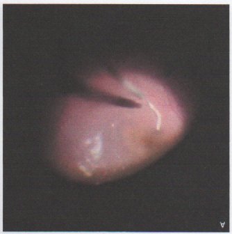
was normal. A picture of the fundiscopic examination is shown below.
DIAGNOSIS: Ocular Gnathostomiasis. On fundoscopy, a worm was seen moving sluggishly
in the posterior segment of his left fundus. A pars plana vitrectomy was performed
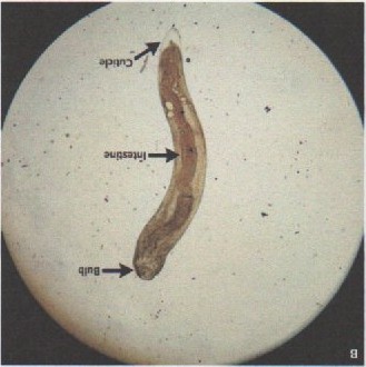
with removal of the worm. Under light microscopy, a larval-stage nematode with a cephalic
bulb, thick cuticle, and well-developed intestine was identified (image on right),
features consistent with Gnathostoma spinigerum. Computed tomography of the brain, orbits, chest, abdomen, and pelvis was unremarkable.
Gnathostomiasis is acquired through ingestion of undercooked freshwater fish, poultry,
snake, or frog in regions where the disease is endemic - the first two of which this
patient from rural central india had consumed. He was treated with oral and ocular
glucocorticoids and a course of albendazole. At the 8-week follow-up, his symptoms
had resolved but his visual acuity remained 20/40 in the left eye owing to the development
of a cataract.